返回首页

肺癌:支气管动脉化疗性栓塞

时间:2020-03-01 18:35来源:未知 作者:admin
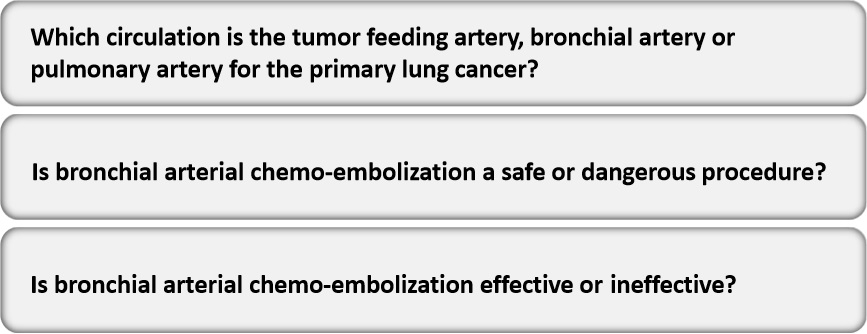
从60,70年代发展起来的这一技术,差不多算是一项古老的技术,曾经被证明对生存率没有影响的技术,几十年过去了,如今它 - Is it logical? - Is it dangerous? - Is it effective? Are there any study is to clarify these interrogative points(疑问点) an
从60,70年代发展起来的这一技术,差不多算是一项古老的技术,曾经被证明对生存率没有影响的技术,几十年过去了,如今它

- Is it logical?
- Is it dangerous?
- Is it effective?
 
Are there any  study is to clarify these interrogative points(疑问点) and to  develop a new treatment method for primary lung cancer.


primary lung cancer
CT Study

Treatment method
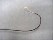
Guiding catheter: 4 Fr. cobra shape, shepherd hook 
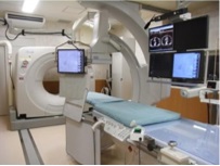
Angio-CT hybrid suits
Chemo-agents & Embolic Material


CT Study
Catheter insertion to the bronchial artery and DSA study
 
Anti-neoplastic agents: CDDP;20-50mg, 5-FU;250-300mg  Embolic material: Hepasphere(100‐150
μm);25mg
 
Actual diameter:200-300 μm
Arterial infusion CT:CM injection: 2.0 – 8.0 mL, 0.2 – 0.4 mL/sec
Chemo-infusion and Embolization;;End point: stasis or slow down of blood flow
CT follow-up:
One month after the treatment
Repetition of treatment:Once a month if effective


Is BACE a logical method ?
Circulation to the pulmonary lesion
 
Feasibility of bronchial arterial catheterization
Degree of Enhancement of the lesion on DSA and arterial infusion CT

Is it dangerous ?
Complications of bronchial chemo-infusion and embolization
Procedure related complications
Spinal arterial circulation

Is it effective ?
Treatment effects
Treatment effect was evaluated after three sessions by CT exams.
RECIST criteria was used to evaluate the treatment effect


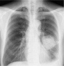
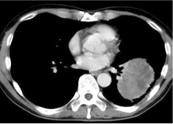
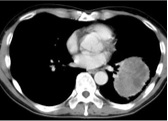
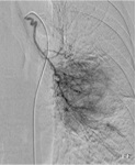
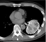
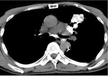
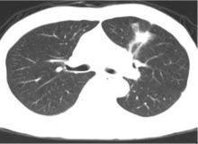
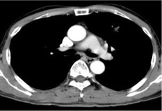
完全染色的病变(Total enhancement of the lesion)
Primary lung cancer:T2N1M1
Poorly deff. adenocarcinoma
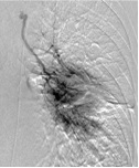
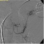
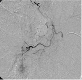
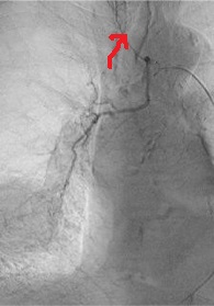
Bronchial angiography  shows tumor brash  corresponding to the  lesion.
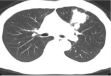
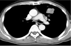
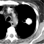
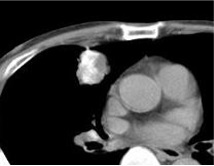
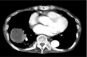
Arterial infusion CT:The pulmonary lesion was  totally enhanced. The lymph  node metastases  was  simultaneously enhanced.
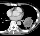
Post BACE (after 3 sessions)
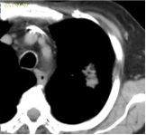
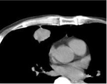
Follow-up CT examination showed
marked regression of the primary lesion.
Respiratory symptom has improved.

Adenocarcinoma Bronchial arteriography
Arterial infusion CT
CT follow-up in 3 months


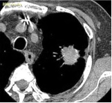
部分染色的病变(Partial enhancement of the lesion)
Primary lung cancer
T2N0M0
Non-small cell lung cancer
 
 
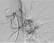
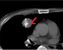
Bronchial angiography  shows tumor brash  corresponding to the  lesion. The pulmonary lesion was  partially  enhanced.The  defect area (→) could be fed  by the other branches.
Post BACE:Treatment effect was SD. The 3ed session:The area treated in the first  session was reduced,  however, the untreated  area (→ ) had progressively  increased .
Ring enhancement of the lesion
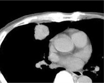
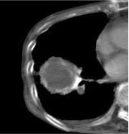
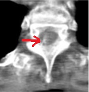
Primary lung cancer:T2N0M0;Bronchial arteriography did not show the enhancement  of the tumor, however, arterial infusion CT demonstrated  a ring and marginal enhancement of the tumor. The intercostal artery was originated from the right bronchial artery ( ) The circulation to the spinal cord was visualized in the arterial infusion CT ( )

Is it logical ?
 primary lung cancer in the lung field is fed by the bronchial  artery. Chemo-infusion into the bronchial artery followed by the embolization is a  logical treatment method.
Is it dangerous ?
There was neither procedure related complication nor chemo-agent related side effects.  The spinal circulation was safely avoided by the use of the arterial infusion CT.
Is it effective ?
The local control rate in three months was 8/9 ( 89%).

Bronchial arterial chemo-embolization is a safe and effective method to control  growth of pulmonary cancer with minimum complications.
 
尽管如此,一个治疗手段的有效性,主要是有质量的寿命年/钱

疗效的判定的包括
近期疗效的判定:客观反应率ORR
远期疗效的判定:OS

COST-EFFECT
 
顶一下
(0)
0%
踩一下
(0)
0%
------分隔线----------------------------
  • 上一篇:没有了
  • 下一篇:没有了
推荐内容